
Facial and Jaw Bone Grafting in Mysore: When Is It Considered?
- Home
- Blog
- About Us
A damaged or deficient area of bone may affect the structure of the jaw or the treatment options available for a patient. Bone grafting is a surgical technique used to add bone material to an area where additional support or reconstruction may be needed. The decision to use a graft depends on the cause and extent of bone loss, the location of the defect and the treatment being planned.
At APTA Clinic in Mysore, bone grafting is listed among the facial surgery services. The clinic’s published information describes its use in situations involving jawbone deficiency, trauma, and bone loss following cyst or tumour removal, as well as in preparation for selected dental implant treatments.
Bone grafting is not necessary for every facial injury or area of bone loss. A clinical assessment is needed to establish whether a graft is appropriate, what material may be used and how the procedure fits into the overall treatment plan.
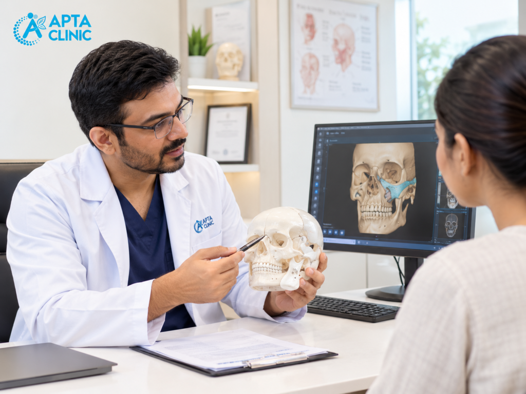
What Is Facial Bone Grafting?
Bone grafting involves placing bone or a suitable bone-graft material in an area where additional bone is required. The material provides a framework or support as the area heals and, depending on the material and circumstances, may integrate with or be replaced by the body’s own bone.
In the face, grafting may involve the jawbone or another facial bone. The purpose depends on the clinical situation. For example, a graft may be considered to rebuild a defect, support a planned reconstruction or provide sufficient bone for a dental implant in a suitable patient.
Bone grafting is a technique, not a diagnosis. It is used for specific clinical reasons and must be planned around the patient’s condition.
Facial trauma refers to an injury affecting the face. It may involve the skin and other soft tissues, the bones of the face, the teeth or structures around the eyes and nose.
Common causes include road traffic accidents, falls, sports injuries and physical assaults. The severity ranges from a superficial wound to a fracture that changes the position of a facial bone or affects normal function.
A facial injury may involve one area or several structures at the same time. For example, a blow to the lower face can injure the jaw and teeth, while an impact around the cheek may affect the cheekbone or the bones surrounding the eye.
The first step is to determine the extent of the injury. Swelling and bruising can make an examination difficult, and a fracture cannot always be ruled out by looking at the face.
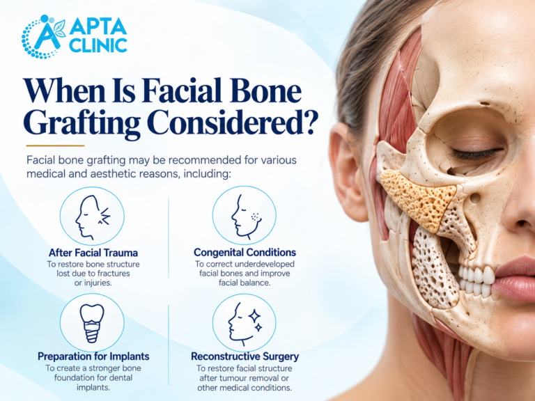
When May Bone Grafting Be Considered?
The need for a graft depends on the amount and location of missing or damaged bone. Several situations may lead a clinician to consider it.
- Bone loss after facial trauma
An accident or other significant injury can damage facial bones. Depending on the type of fracture and the extent of bone loss, treatment may involve fracture repair, stabilisation or reconstruction.
A bone graft may be considered when a defect requires additional bone to support reconstruction. It is not required for every facial fracture. The decision depends on the injury, the remaining bone, the condition of nearby tissues and the overall treatment plan.
A recent serious facial injury needs prompt medical assessment. Difficulty breathing, uncontrolled bleeding, loss of consciousness, major vision changes or a suspected head or neck injury require emergency care rather than a routine appointment.
- Bone defects following cyst or tumour treatment
Some jaw cysts and tumours can affect the surrounding bone. When a lesion is removed, the resulting defect may need monitoring or further treatment.
In selected cases, bone reconstruction may be considered to address a significant defect. The decision depends on the nature of the lesion, the amount of bone removed, the condition of the surrounding tissues and the patient’s recovery.
The underlying lesion must be assessed and managed appropriately first. A graft is not a substitute for diagnosing the cause of a jaw lesion.
- Insufficient jawbone before dental implants
Dental implants require suitable bone at the planned placement site. If the jawbone does not provide enough volume or has an unfavourable shape, a clinician may consider bone augmentation before or alongside implant treatment, depending on the case.
The amount and shape of available bone, the position of nearby structures, gum health and the overall dental plan all influence the decision.
Bone grafting does not automatically make every patient suitable for implants. The clinician should explain whether a graft is likely to help, whether alternatives exist and what additional stages of treatment may be required.
- Certain congenital or structural bone defects
Some patients have a bone defect related to a congenital condition or a previous operation. In selected cases, reconstruction may involve bone grafting as part of a broader treatment plan.
The approach varies with the location and size of the defect, age, development, previous treatment and the function that needs to be addressed. These cases may require coordination between different specialists.
- Bone loss related to disease or previous treatment
Certain diseases, infections or previous surgical procedures can leave areas of deficient bone. The cause must be assessed before reconstruction is planned.
The treating clinician may need to address the underlying problem and establish that the site is suitable for grafting. Not every area of bone loss benefits from a graft, and the decision should be based on clinical findings rather than appearance alone.
What Types of Bone Graft Materials Are Used?
The material used depends on the treatment goal, the amount of bone needed and the patient’s circumstances. Common categories include the following.
Bone from the patient’s own body
This is called an autograft. Bone is taken from another site in the same patient and transferred to the area that needs reconstruction.
The suitability of this approach depends on the amount of bone required and the donor site. Taking bone from another part of the body may involve an additional surgical site and its own recovery considerations.
Donor bone
Donor bone, known as an allograft, comes from another human donor and is processed for medical use. Whether it is appropriate depends on the procedure, the available material and the treating clinician’s judgement.
Synthetic or other bone-graft materials
Some procedures use manufactured materials or other suitable graft substitutes. These materials differ in their composition and behaviour, so they are not interchangeable in every situation.
The treating clinician should explain which type of material is being proposed, why it suits the case and whether other options are available. A patient should not assume that one material is best for every facial or jaw reconstruction.
How Is Bone Grafting Planned?
Planning begins with understanding the reason for the bone defect and what the treatment is intended to achieve.
Medical and dental history
The clinician may ask about previous injuries or operations, medical conditions, current medicines, allergies, smoking or tobacco use and any history of infection or delayed healing. These details can influence surgical planning.
Examination of the affected area
The examination depends on the location of the defect. It may involve the jaw, teeth, gums, facial structure and surrounding tissues.
Imaging and investigations
Dental X-rays or other imaging may be used to assess the amount and shape of available bone. A CT scan or other investigation may be appropriate in selected cases. The clinician determines which tests are needed for the specific treatment plan.
Discussion of options
The clinician explains whether grafting is indicated, what material may be used, whether treatment will require more than one stage and what alternatives are available.
Patients should also understand the likely recovery period, possible complications, follow-up requirements and estimated costs before consenting to the procedure.
What Happens During a Bone Grafting Procedure?
The exact steps vary with the location of the defect, the amount of bone required and the type of graft material.
In general, the surgical team accesses the affected area, prepares the site and places the selected graft material. The graft may need to be secured, and the surrounding tissue is closed or managed according to the procedure.
The type of anaesthesia, the setting in which treatment takes place and whether the patient can go home the same day depend on the planned operation and individual circumstances.
Some patients need additional treatment before the next stage can proceed. For example, bone augmentation before dental implants may require a healing period and reassessment before implant placement.
Ask the treating clinician to explain the planned steps for your procedure rather than relying on a general description of bone grafting.
Recovery After Bone Grafting
Recovery depends on the location of the graft, the extent of surgery, the material used and the patient’s overall health. A smaller graft in the jaw may involve different aftercare from a more extensive facial reconstruction.
The treating team may provide instructions about:
- Wound care and oral hygiene
- Medicines prescribed for pain or other post-operative needs
- Food texture and chewing, where relevant
- Activity restrictions and avoiding pressure or impact to the treated area
- Follow-up appointments and any required imaging
- When further treatment, such as dental implant placement, can be considered
Bone healing and integration take time. Improvement in swelling or discomfort does not necessarily mean that the graft is ready for the next treatment stage.
Attend follow-up appointments and follow the instructions provided. Contact the treating team if pain or swelling worsens, you develop a fever, the wound opens or there is discharge or another unexpected change. Severe symptoms require urgent medical assessment.
Risks and Limitations of Bone Grafting
Bone grafting is a surgical procedure and has potential risks. Depending on the case, these may include infection, bleeding, swelling, pain, delayed healing, graft loss or failure to achieve the intended amount of bone. If bone is taken from another part of the patient’s body, that donor site may have its own complications.
The outcome also depends on the original defect, the health of the surrounding tissues, the treatment method and individual healing factors. A graft cannot guarantee that a later procedure, such as dental implant placement, will be possible.
Before proceeding, ask the clinician about the risks that apply to your situation, how healing will be assessed and what alternatives are available if grafting is not suitable or does not achieve the intended result.
How Much Does Bone Grafting Cost in Mysore?
The cost depends on the location and size of the defect, the type and quantity of graft material, investigations, surgical complexity, the facility where treatment is performed and the follow-up required.
If grafting is part of a larger treatment plan, ask whether the estimate includes the consultation, imaging, procedure, graft material, medicines, follow-up and any later treatment.
APTA Clinic’s current procedure-specific fees are not confirmed here. Contact the clinic for its consultation charges and a treatment estimate after assessment. Insurance coverage or reimbursement, if relevant, should be checked with the provider and insurer before treatment.
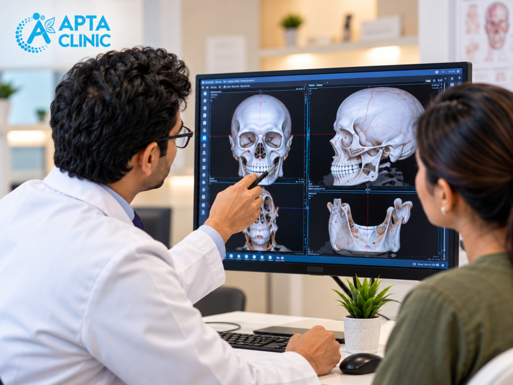
Prepare for a consultation
If you have already undergone an examination or procedure, bring available scan reports, X-rays, treatment records and a list of current medicines. If a dentist or another clinician has recommended grafting, bring that referral or treatment plan if available.
Useful questions to ask include:
- Why is bone grafting being considered in my case?
- What happens if I do not have the graft?
- Which graft material is proposed, and why?
- Will the treatment require more than one procedure?
- What risks and recovery restrictions should I expect?
- How will the clinician check whether the graft has healed?
- What is included in the cost estimate?
What Happens if a Bone Graft Does Not Integrate Properly?
Bone graft integration is an important part of the healing process. After placement, the graft needs suitable surrounding tissue conditions and adequate stability to support the intended reconstruction. In some cases, the graft may not integrate as expected, or part of the graft material may be lost during healing. This can affect the amount of bone available for the next stage of treatment.
Several factors may contribute to unsuccessful healing, including infection, wound breakdown, movement at the graft site and individual health-related factors. The significance of these problems depends on the procedure and the extent of the affected area. A graft that does not integrate successfully does not always mean that further reconstruction is impossible, but it may require reassessment and a revised treatment plan.
The treating surgeon may recommend clinical examination or imaging to evaluate the site before deciding on the next steps. Patients should report increasing pain, persistent swelling, wound opening, unusual discharge or exposed graft material rather than attempting to manage these problems themselves.
Questions to Ask Before Facial Bone Grafting
A consultation is an opportunity to understand the purpose of the procedure and make an informed decision. Patients may wish to ask what caused the bone deficiency, whether grafting is necessary and what the proposed procedure is expected to accomplish.
It is also useful to discuss the type of graft material, whether bone will be harvested from another site, what imaging or investigations are needed and whether another procedure will be required later. Ask about the likely recovery period, activity restrictions, follow-up schedule and the risks that are most relevant to your situation.
Patients should also understand the alternatives, including the option of delaying or not proceeding with surgery where clinically appropriate. The decision should be based on clear information about the expected benefits, limitations and risks.
Facial Bone Grafting in Mysore: Getting the Right Assessment
People searching for facial bone grafting in Mysore may have different concerns, including bone loss after tooth extraction, a facial injury, a jaw defect or a structural difference affecting the face. These conditions do not all require the same treatment, and a general description cannot determine whether grafting is suitable for an individual patient.
How Do Specialists Check Whether a Bone Graft Has Healed?
Assessing bone graft healing involves more than checking whether the surgical wound has closed. The surrounding soft tissues may heal before the underlying bone has completed its integration and remodelling. For this reason, the surgeon considers both the condition of the surgical site and the progress of the reconstructed area.
Clinical reviews may include checking for tenderness, swelling, wound healing and signs of infection. Depending on the purpose of the graft, imaging may be used to assess bone formation, graft position and the amount of bone available for further treatment. The type and timing of imaging depend on the procedure and the clinical question being investigated.
There is no single healing timeline that applies to every bone graft. The size of the defect, graft material, surgical technique and individual healing response can all influence the assessment. The treating clinician determines when the graft is ready for the next stage of treatment, rather than relying on a fixed number of weeks or months.
Can a Bone Graft Change Shape During Healing?
Bone grafts can undergo changes as the surrounding tissues heal and the graft remodels. Depending on the material and procedure, some reduction in graft volume may occur over time. The extent of this change varies, and it does not automatically mean that the treatment has failed.
The location of the graft, the size of the original defect and the demands placed on the reconstructed area can influence the outcome. In facial reconstruction, the surgeon considers whether the graft provides the required structural support. In dental procedures, the available bone volume may also influence whether a planned implant or other restoration can proceed.
Follow-up examinations help determine whether the graft is maintaining sufficient volume for its intended purpose. If the result differs from the treatment plan, the specialist can assess whether observation, additional treatment or a different approach is appropriate.
How Do Smoking and Daily Habits Affect Bone Graft Healing?
Bone healing depends on several biological processes, including adequate blood supply and healthy surrounding tissues. Smoking can interfere with these processes and is associated with a higher risk of complications in many surgical settings. Patients who smoke should discuss this with their surgeon before undergoing bone grafting and ask about an appropriate plan for stopping or reducing exposure around the procedure.
Daily habits may also affect recovery. Poor oral hygiene can increase the risk of complications following grafting procedures inside the mouth, while activities that place pressure on a healing site may interfere with the surgeon’s instructions. Patients should follow the recommended cleaning routine, dietary guidance and activity restrictions for their particular procedure.
Nutrition and management of existing health conditions are also relevant to overall healing. Patients should tell their healthcare provider about prescribed medicines, supplements and medical conditions before surgery. Medication changes should only be made with guidance from the relevant clinician.
What Is the Difference Between Bone Graft Healing and Bone Remodelling?
Bone graft healing and bone remodelling are related but distinct processes. Healing involves the early response of the tissues following surgery, including wound closure and the biological processes that allow the graft to interact with the surrounding area. Remodelling is the longer-term process through which bone tissue changes and adapts over time.
The speed and extent of these processes depend on the type of graft, the surgical site and individual biological factors. A wound that appears to have healed does not necessarily indicate that the reconstructed bone has completed remodelling. Likewise, the absence of pain alone cannot confirm that the graft has integrated adequately.
Understanding this distinction helps explain why follow-up may continue after the initial recovery period. The surgeon uses clinical findings and, when appropriate, imaging to decide whether the reconstructed area is progressing as expected and whether further treatment can safely proceed.
Book a Bone Grafting Consultation in Mysore
If you have been told that you may need jawbone reconstruction or bone grafting, an assessment can help clarify the reason for treatment and the options available.
Contact APTA Clinic to ask about an appointment and confirm whether the clinic can assess your specific concern.
APTA Clinic
- Location: Krishnamurthy Puram, Mysore, Karnataka
- Phone: +91 7411 707 555
- Website: https://aptaclinic.com/
- Appointment enquiries: https://aptaclinic.com/contact/
Confirm the current address, consultation availability and service details before visiting.
Frequently Asked Questions (FAQs)
Facial bone grafting is a surgical technique used to add bone or suitable graft material to an area where additional bone is needed. It may be considered for selected jaw or facial defects, depending on the cause and extent of bone loss.
A clinician may consider it when the jaw lacks sufficient bone for a planned dental implant, when a defect remains after trauma or surgery, or when reconstruction is needed for a specific condition. Not every patient with bone loss requires a graft.
No. The need depends on the amount and shape of available jawbone and the planned implant position. A dental clinician assesses whether the existing bone is sufficient or whether augmentation should be considered.
Depending on the procedure, the material may come from the patient’s own body, a human donor or a manufactured bone-graft substitute. The appropriate option depends on the defect, treatment goal and clinical assessment.
Healing varies with the size and location of the graft, the procedure and individual factors. The treating clinician will advise when the area should be reviewed and whether additional treatment can proceed.
The procedure is performed with an appropriate anaesthetic plan, but discomfort, swelling and tenderness can occur during recovery. The amount and duration vary. Ask the treating clinician what to expect and how post-operative discomfort will be managed.
Potential risks include infection, bleeding, swelling, delayed healing and problems with graft integration. If bone is taken from another part of the body, the donor site can also have complications. The risks specific to the proposed procedure should be discussed before consent.
It may be considered for selected defects after an injury. Treatment depends on the fracture, the remaining bone and surrounding tissues, and whether reconstruction is needed. Recent serious injuries should be assessed urgently.
APTA Clinic lists locations in Krishnamurthy Puram and Vijayanagar, Mysore.