
White Patches on the Skin: Vitiligo, Fungal Infection or Something Else?
- Home
- Blog
- About Us
Finding a white or noticeably lighter patch on your skin can raise an immediate question: is it vitiligo, a fungal infection or something else?
Colour alone cannot reliably answer that question.
Several skin conditions can cause areas of lighter pigmentation. Vitiligo is one possibility, but tinea versicolor, changes following inflammation or injury, and other pigment disorders can look similar. An accurate diagnosis helps determine whether treatment is needed and which approach is appropriate.
If you are searching for vitiligo treatment in Mysore, the first step is a dermatological assessment rather than assuming what the patch represents.
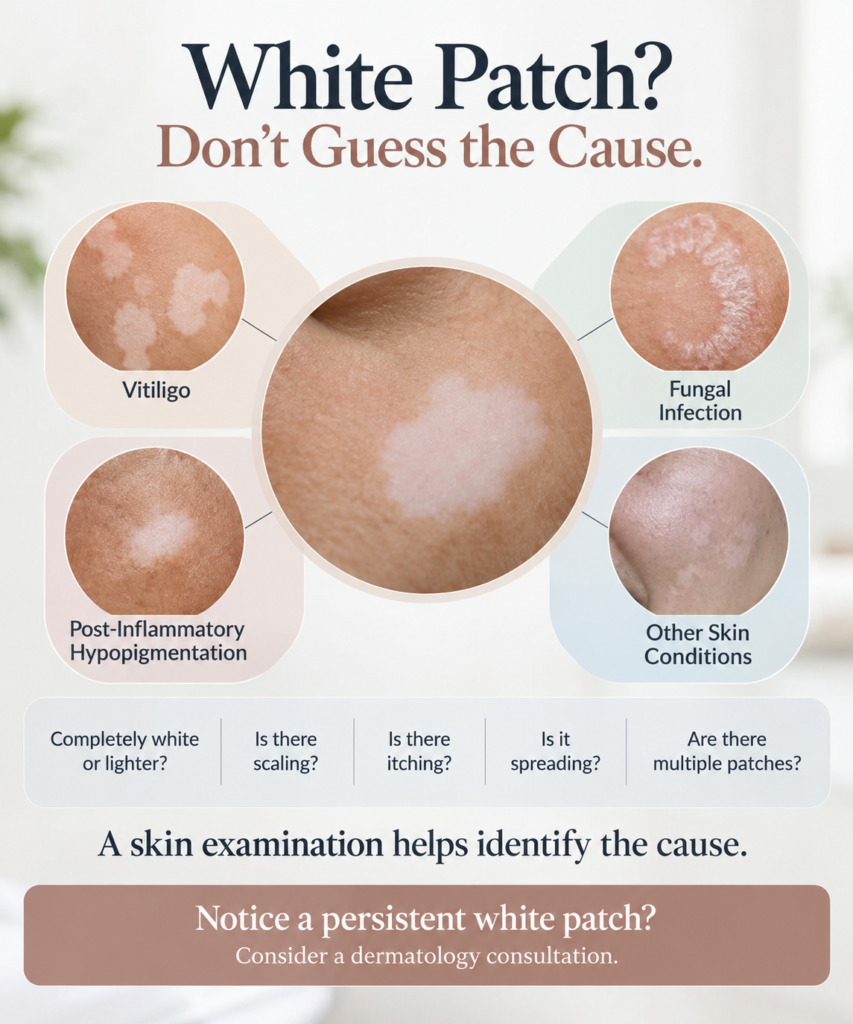
What Can White Patches on the Skin Mean?
White or lighter patches occur when an area of skin has less pigment than the surrounding skin.
Possible causes include:
- Vitiligo
- Fungal infections such as tinea versicolor
- Pigment changes after inflammation
- Skin changes following an injury or irritation
- Other pigmentation disorders
A dermatologist will usually consider more than the colour of the patch. The location, surface, border, duration and changes over time can all provide useful information.
The doctor may also ask:
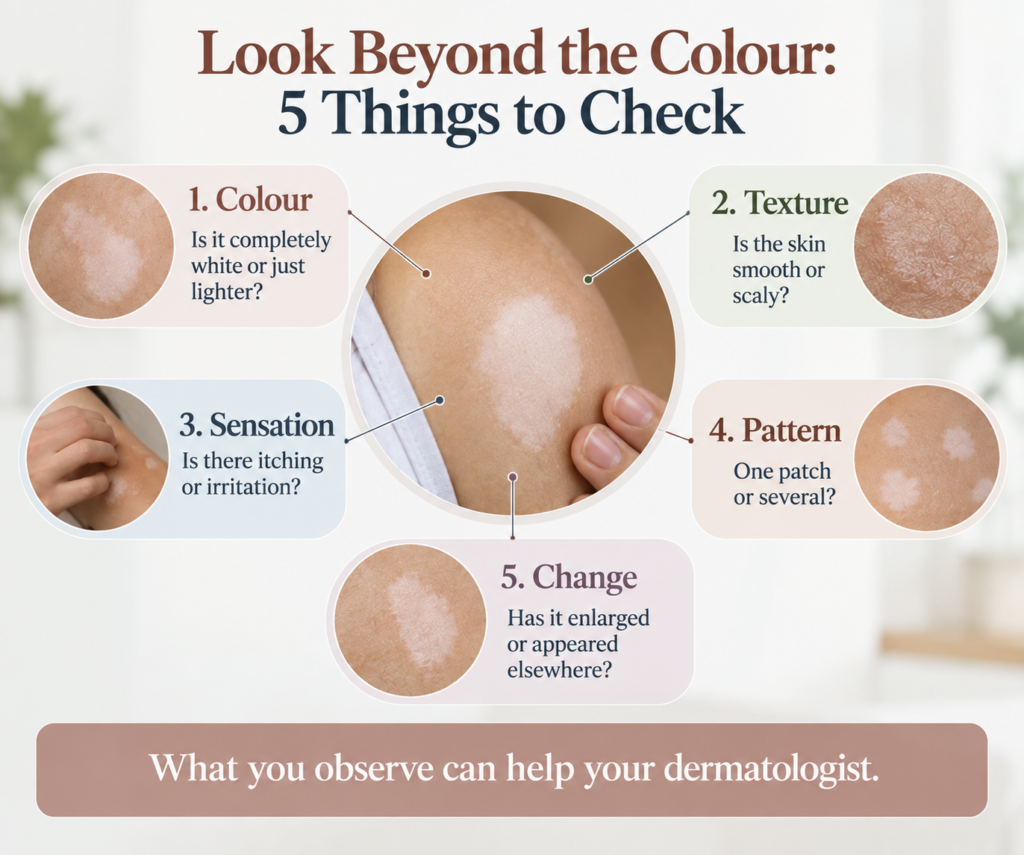
- Is the patch completely white or simply lighter?
- Is the border clearly defined?
- Is the surface smooth or scaly?
- Does it itch?
- How long has it been present?
- Is it becoming larger?
- Are new patches appearing?
- Has hair within the affected area changed colour?
These details can help with the assessment, but they cannot be used as a reliable substitute for diagnosis.
What Is Vitiligo?
Vitiligo is a condition in which areas of skin lose their normal colour because melanocytes, the cells responsible for producing pigment, are affected.
The patches may initially appear lighter than the surrounding skin and can become completely white. Vitiligo can occur on different parts of the body, including the face, hands, feet and areas around body openings. Hair within an affected area can also become white or grey.
Vitiligo is not a fungal infection and is not contagious.
What Do Vitiligo Patches Look Like?
Vitiligo can look different from one person to another.
The patches may:
- appear lighter than the surrounding skin
- become completely white
- have noticeable borders
- occur in more than one area
- increase in size over time
- occur on both sides of the body in some forms
- cause hair in the affected area to become white or grey
Some people experience itching when vitiligo first appears or becomes active, although significant discomfort is not typical.
These features can suggest vitiligo, but they do not confirm it. A dermatologist may use examination and additional tools when necessary.
What Is Tinea Versicolor?
Tinea versicolor is a common skin condition caused by an overgrowth of yeast that normally lives on the skin.
It can produce patches that are lighter or darker than the surrounding skin. The affected areas may be dry or scaly and can sometimes itch. The patches commonly occur on the chest, back, shoulders and other areas of the trunk.
Because tinea versicolor can produce lighter patches, it is sometimes confused with vitiligo.
The two conditions have different causes and require different management.
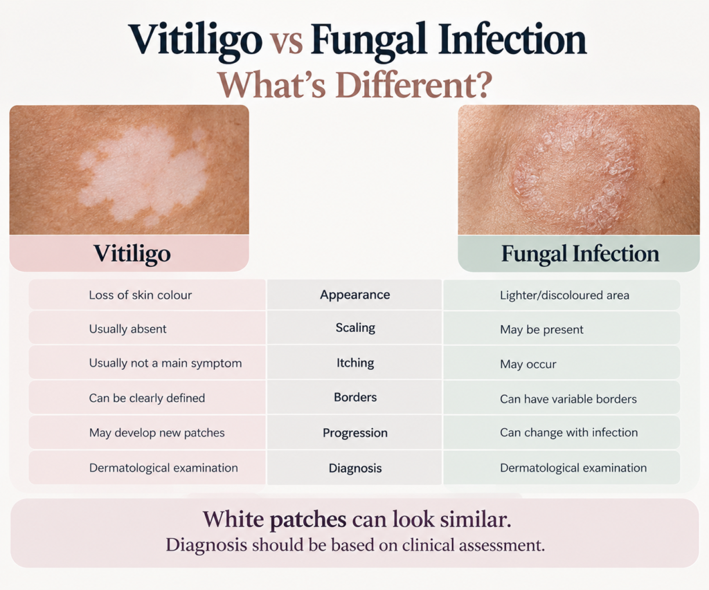
Vitiligo vs Fungal Infection: Are There Any Clues?
There can be clues, but appearance alone should not be treated as a diagnosis.
| FEATURE | VITILIGO | Tinea versicolor |
|---|---|---|
|
Main change |
Loss of skin pigment |
Skin colour change associated with yeast overgrowth |
|
Colour |
Often clearly lighter or white |
Can be lighter or darker |
|
Surface |
Usually smooth rather than scaly |
May be dry or scaly |
|
Itching |
May occasionally itch |
Can itch |
|
Contagious |
No |
Not generally considered contagious |
Tinea versicolor commonly produces fine scaling, while vitiligo primarily involves loss of pigment rather than a scaly surface. However, skin conditions do not always follow a textbook appearance.
Can Itching or Scaling Tell You What the Patch Is?
Itching and scaling can provide useful clues, but neither confirms the diagnosis.
Tinea versicolor may produce dry or scaly patches and can cause itching. Vitiligo usually does not cause significant discomfort, although some people may experience itching when patches first appear or become active.
A white patch that itches is therefore not automatically a fungal infection. Likewise, a smooth white patch should not automatically be labelled as vitiligo.
What Other Conditions Can Cause Lighter Skin?
Vitiligo and fungal infections are not the only possible explanations.
Skin may become lighter after inflammation, irritation or an earlier skin problem. Certain injuries can also leave changes in pigmentation. The AAD notes that several conditions can cause loss of skin colour and that accurate diagnosis is important before treatment is selected.
The dermatologist may consider:
- your skin type
- where the patch is located
- previous inflammation or rashes
- injury or irritation
- how long the patch has been present
- whether it is changing
- associated symptoms
- other relevant medical history
This is why a new pigment change should be assessed in context rather than identified from a photograph alone.
Why Do Location and Progression Matter?
Two patches can look similar while having different causes.
Vitiligo can affect the face, hands, feet, fingers, wrists and areas around the eyes and mouth. Hair within affected areas can also lose its colour.
Tinea versicolor commonly affects the trunk and may produce fine scaling.
The history also matters. A lighter patch that developed after an earlier rash or inflammation may have a different explanation from one that appeared without any preceding skin problem.
When Should You See a Dermatologist for White Patches?
Consider arranging a dermatology consultation when:
- a new white or lighter patch appears without an obvious explanation
- the patch is getting larger
- additional patches are appearing
- the colour change persists
- the area is itchy or scaly
- there is redness or inflammation
- hair within the affected area becomes white or grey
- home treatment has not helped
- you are unsure whether the patch is fungal, vitiligo or another condition
You do not need to wait until several patches appear before asking a dermatologist about an unexplained pigment change.
The purpose of the consultation is to establish what the skin change represents and whether treatment or monitoring is appropriate.
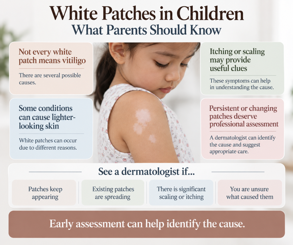
Why Can Self-Diagnosing White Patches Be Misleading?
Online photographs can be useful for general education, but they have limitations.
Lighting, tanning, camera exposure and image quality can all change how skin appears. More importantly, several conditions can produce similar-looking changes.
The AAD specifically recommends obtaining an accurate diagnosis when skin develops lighter or white patches because the cause may be vitiligo, tinea versicolor, a skin injury or another condition.
Using an antifungal treatment simply because a patch looks white may be inappropriate if the underlying problem is not fungal.
Similarly, assuming that every white patch is vitiligo can cause unnecessary concern.
How Does a Dermatologist Evaluate White Patches?
The assessment generally begins with your medical history and an examination of the affected skin.
Your dermatologist may ask:
- When did you first notice the patch?
- Has it changed in size?
- Have other patches appeared?
- Does it itch?
- Is the surface scaly?
- Was there a rash or injury in the same area before the colour changed?
- Have you applied any creams?
- Do you have other skin conditions?
- Does anyone in your family have similar pigment changes?
The doctor may then examine the area under appropriate lighting.
Further testing depends on what the examination shows.
Wood's Lamp Examination
A Wood’s lamp is a specialised light that can make certain areas of pigment loss easier to see.
The American Academy of Dermatology notes that dermatologists may use a Wood’s lamp when evaluating suspected vitiligo.
It can also be used in some cases when tinea versicolor is suspected.
Skin Examination or Scraping
If a fungal infection such as tinea versicolor is suspected and the diagnosis is uncertain, a dermatologist may take a small skin scraping and examine it under a microscope.
Other Tests
Additional testing is not necessary for every person with a white patch.
Depending on the clinical findings, evaluation of suspected vitiligo can sometimes include a skin biopsy or blood tests. Mayo Clinic notes that these tests may be used selectively as part of the diagnostic process.
The appropriate evaluation depends on the individual situation.
Does Every White Patch Need Treatment?
No.
Treatment depends on the underlying diagnosis and how the condition is affecting the individual.
A fungal infection requires treatment directed at the infection. Vitiligo has different management options. Some pigment changes may simply need observation.
The important step is to establish the cause before deciding what should be applied to the skin.
What Treatments May Be Used for Vitiligo?
There is no single treatment that suits everyone with vitiligo.
Treatment decisions can depend on:
- age
- location of the affected skin
- extent of pigment loss
- whether the condition is progressing
- previous treatment
- overall health
- individual preferences
Depending on the situation, treatment may include topical medicines, light-based treatment, camouflage and selected surgical or cellular approaches. The AAD and Mayo Clinic both note that treatment selection depends on the individual case and that results can vary.
Treatment may take time, and follow-up can be necessary.
Why Is Sun Protection Important With Vitiligo?
Areas affected by vitiligo contain less melanin and can be more vulnerable to sunburn.
Protecting depigmented skin from excessive ultraviolet exposure is therefore important. Mayo Clinic recommends broad-spectrum sunscreen with an SPF of at least 30, along with measures such as seeking shade and using protective clothing.
Your dermatologist can advise you on sun protection that suits your skin and daily routine.
What About White Patches Caused by a Fungal Infection?
When the diagnosis is tinea versicolor, treatment is directed at the fungal infection.
The choice of treatment can depend on factors such as the location and extent of the affected skin.
An important point is that skin colour may remain uneven even after the fungal infection has been treated. The lighter appearance can take time to return to its usual colour.
Therefore, lighter skin after treatment does not necessarily mean that the infection is still active.
A dermatologist can assess whether the infection has cleared and whether the remaining colour difference requires further management.
Common Misconceptions About Vitiligo
1. “Every white patch is vitiligo.”
No.
Vitiligo is one possible cause. Tinea versicolor, post-inflammatory changes, injury and other pigment disorders can also produce lighter skin.
2.“Vitiligo is a fungal infection.”
No.
Vitiligo involves loss of skin pigment and is not a fungal infection.
3.“Vitiligo spreads from one person to another.”
No.
Vitiligo is not contagious. It cannot be transmitted through ordinary contact.
4.“Every white patch needs treatment.”
Not necessarily.
Treatment depends on the diagnosis and the individual’s circumstances.
5.“Vitiligo can be diagnosed from a photograph.”
A photograph may provide a useful reference, but it cannot reliably replace an appropriate clinical examination.
What Should You Ask During a Dermatology Consultation?
If you have noticed white or lighter patches, consider asking:
- What could be causing this colour change?
- Does the patch look like loss of pigment or another skin condition?
- Would a Wood’s lamp examination be useful?
- Could a previous rash or injury explain the colour change?
- If this is vitiligo, does it appear active or stable?
- If this is fungal, how will we know whether the infection has cleared?
- What treatment options are appropriate for my diagnosis?
- Do I need specific sun protection?
- How should I monitor the patch?
- When should I return for review?
Taking photographs over time can also help document whether the size or appearance of the patch is changing.
Vitiligo Treatment in Mysore: Why Diagnosis Comes First
If you are searching for vitiligo treatment in Mysore, diagnosis should come before choosing a treatment.
The information supplied for this page states that APTA Clinic provides dermatology services and identifies Dr. Swathi Anand as a Dermatologist & Aesthetician.
APTA’s current website identifies Dr. Swathi Anand as an MBBS, DDVL dermatologist and aesthetician and lists her dermatology and aesthetic practice at APTA Clinic, Mysuru.
The clinic currently lists branches in Krishnamurthy Puram and Vijayanagar, Mysore, with the phone number +91 7411 707 555.
The supplied page also discusses clinical examination, Wood’s lamp examination and, when required, investigations such as biopsy or blood tests. It mentions treatment approaches including topical medicines and narrowband UVB phototherapy.
These options should not be presented as suitable for every person with a white patch. The appropriate treatment depends on the diagnosis and individual clinical findings.
The Main Point
A white patch on the skin does not automatically mean vitiligo.
It may be caused by a fungal infection, a change following inflammation or injury, or another pigmentation disorder. Some patches itch or scale, while others cause no discomfort.
The useful first step is to determine why the skin has changed colour.
If a white or lighter patch persists, becomes larger, keeps returning or is difficult to explain, a dermatologist can examine it and decide whether further evaluation is needed.
Once the cause is established, treatment or monitoring can be discussed according to the individual’s situation.
Frequently Asked Questions (FAQs)
No. White or lighter patches can have several causes, including vitiligo, tinea versicolor, post-inflammatory pigment changes and other skin conditions. An accurate diagnosis is important before treatment is selected.
There can be clues, but appearance alone cannot reliably establish the diagnosis. Tinea versicolor may cause lighter or darker patches with fine scaling and sometimes itching. Vitiligo primarily involves loss of skin pigment and generally does not produce scaling.
Yes. Tinea versicolor can produce lighter-looking patches, particularly on the trunk. The affected skin may also be dry or scaly.
It is reasonable to seek dermatological evaluation when a white patch is new, persistent, enlarging, recurring or unexplained, particularly if additional patches appear or the area develops other changes.
A Wood’s lamp is a specialised light used during examination of certain skin conditions. It can make areas of pigment loss easier to see and may assist in evaluating suspected vitiligo.
Depending on the individual situation, options can include topical medicines, phototherapy, camouflage and selected surgical or cellular approaches. Treatment depends on factors such as the location and extent of pigment loss and whether the condition is progressing.
APTA Clinic lists dermatology among its services, and its website identifies Dr. Swathi Anand as a Dermatologist & Aesthetician.
APTA Clinic currently lists locations in Krishnamurthy Puram and Vijayanagar, Mysore. Its published contact number is +91 7411 707 555. Confirm current appointment availability directly with the clinic.
It is better not to assume the diagnosis from appearance alone. White patches can have several causes, so treatment should follow appropriate evaluation when the diagnosis is uncertain.
Note when the patch first appeared, whether it has changed, whether there was a previous rash or injury in the same area, and which creams or medicines you have used. Photographs taken over time may also help show how the patch has changed.