
Mole Removal in Mysore: When Is Removal Recommended and What Should You Expect?
- Home
- Blog
- About Us
Most moles are harmless and do not need treatment. However, a mole that changes in size, shape or colour, begins bleeding or becomes persistently itchy should be assessed by a healthcare professional.
Mole removal may be considered for medical reasons, repeated irritation or cosmetic concerns. Before deciding on a procedure, the first step is to establish what the growth is and whether it needs further investigation.
If you are researching mole removal in Mysore, understanding the assessment process, available removal methods, recovery and possible scarring can help you prepare for a consultation. A mole that looks suspicious should not be treated as a routine cosmetic concern because it may require a biopsy or laboratory examination.
APTA Clinic is described in the supplied information as an aesthetic clinic in Mysore with a dermatology department. Contact the clinic to confirm whether assessment and removal of your particular mole are currently available.
What Is a Mole?
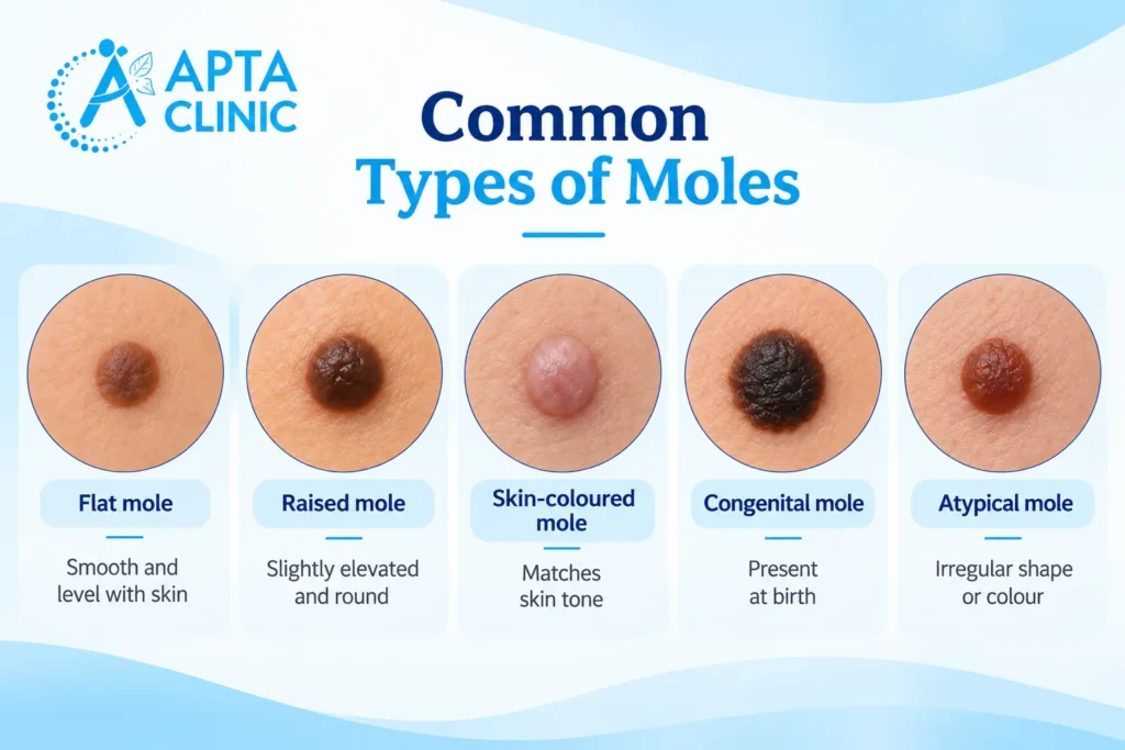
A mole, also called a melanocytic naevus, is a skin growth formed by clusters of pigment-producing cells. Moles can be flat or raised and may appear brown, black, pink, skin-coloured or other shades. Some are present at birth, while others develop during childhood or adulthood.
Many moles remain stable for years. Some change gradually over time, and certain changes may not be dangerous. However, a new or changing mole should not automatically be assumed to be harmless.
Most moles do not require removal. Depending on the mole’s appearance, symptoms, location and medical history, a healthcare professional may recommend monitoring, further assessment or removal.
When Is Mole Removal Recommended?
Mole removal may be considered for several reasons. The recommended approach depends on whether the mole appears medically concerning, causes repeated irritation or is being considered for cosmetic reasons.
- A mole has suspicious changes
A mole that changes in size, shape or colour, develops an irregular border, or starts bleeding, crusting or itching needs professional assessment.
These features do not automatically mean that the mole is cancerous, but they should not be ignored. If a clinician suspects a skin cancer such as melanoma, further investigation or removal for laboratory examination may be necessary.
In this situation, the priority is obtaining an accurate diagnosis rather than improving the mole’s appearance.
- A mole is repeatedly irritated
A raised mole may catch on clothing, jewellery or a razor. Repeated friction can make it uncomfortable or cause occasional bleeding from injury.
A clinician can assess whether removal is appropriate and explain the potential risks, including scarring, infection and bleeding.
- A mole causes cosmetic concern
Some people wish to remove a stable mole because they dislike its appearance or feel self-conscious about it. Cosmetic removal may be an option after the mole has been assessed and considered suitable.
However, a cosmetic request should not replace medical evaluation. If the mole has suspicious features, the clinician should determine whether diagnostic testing is needed before deciding on a removal method.
- The diagnosis is uncertain
Some skin growths resemble moles but are actually other conditions. If a lesion has an unusual appearance or its diagnosis is uncertain, a clinician may recommend further assessment or a biopsy.
Do not try to remove an unidentified growth at home. Cutting, burning or applying unverified mole-removal products can cause injury, infection and scarring. It may also delay the diagnosis of a serious condition.
How to Recognise Warning Signs in a Mole
The ABCDE guide helps identify common warning signs associated with melanoma. It is not a diagnostic test, and a mole can require assessment even if it does not show every feature.
- A – Asymmetry: One half looks different from the other.
- B – Border: The edge is irregular, ragged, scalloped or poorly defined.
- C – Colour: The mole has several colours or an uneven distribution of colour.
- D – Diameter: The mole is larger than about 6 millimetres, although melanoma can be smaller.
- E – Evolving: The mole changes in size, shape, colour or texture over time.
Other warning signs include persistent itching, bleeding, crusting, pain or a sore that does not heal. A new or unusual spot that looks different from your other moles should also be assessed.
These signs do not confirm cancer. However, if you notice them, arrange a medical assessment promptly rather than booking cosmetic removal without an examination.
What Are the Options for Mole Removal?
The appropriate method depends on the mole’s appearance, location, size, depth and whether the tissue needs to be examined in a laboratory.
The following methods are used in clinical practice. Their inclusion here does not confirm that each procedure is offered at APTA Clinic.
- Surgical excision
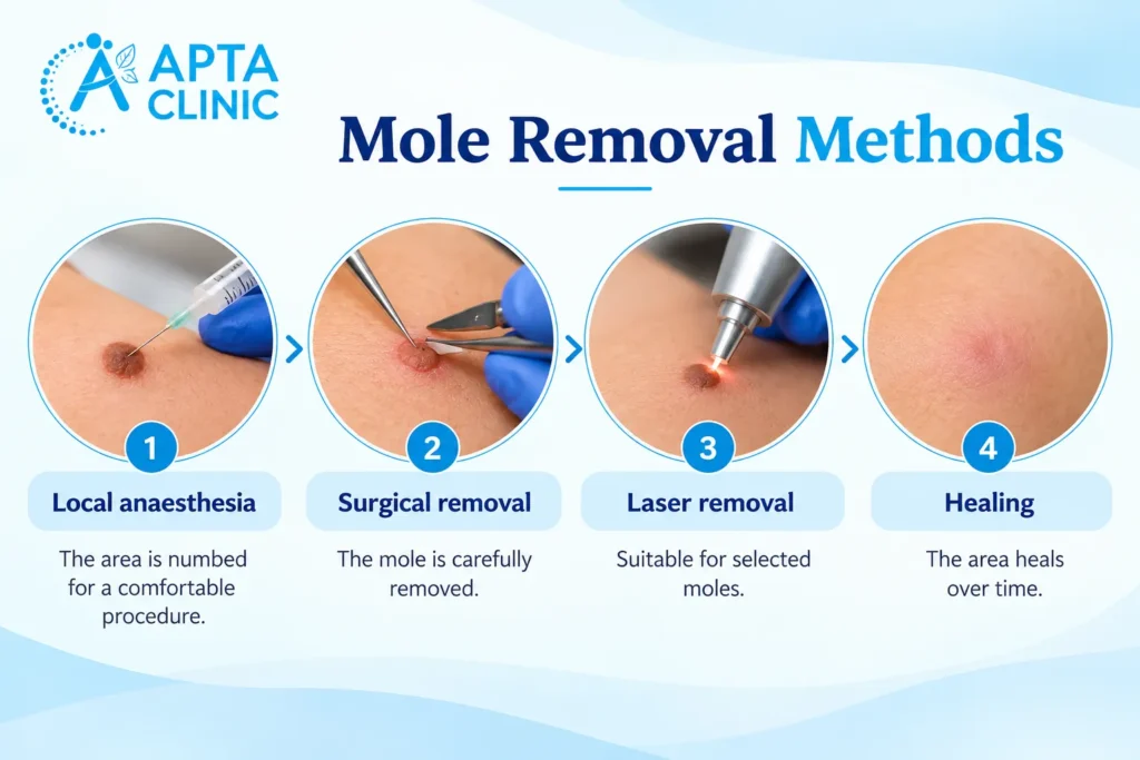
Surgical excision involves removing the mole using a cutting procedure. It may be recommended when a lesion needs to be removed for diagnostic purposes or when its characteristics make excision appropriate.
Depending on the case, the removed tissue may be sent for histopathological examination. This involves a specialist examining the tissue under a microscope to help establish the diagnosis.
Excision creates a wound and may leave a scar. The scar’s size and appearance depend on factors such as the mole’s size and location, the technique used and individual healing.
- Shave removal
For selected raised lesions, a clinician may consider a shave procedure that removes tissue closer to the skin surface.
Whether this method is appropriate depends on the type of lesion and whether a deeper or complete specimen is needed for diagnosis. Shave removal is not suitable for every mole.
If a mole has suspicious features, the removal method must allow the lesion to be assessed appropriately.
- Other removal techniques
Other techniques may be considered for selected skin lesions, depending on the diagnosis. The important question is not simply which procedure is quickest, but whether it is appropriate for the specific growth.
Do not assume that laser treatment, cautery or another cosmetic procedure is suitable for a mole without medical assessment. Some techniques may not provide an adequate tissue sample when laboratory examination is needed.
Your clinician should explain the reason for the recommended method and whether the removed tissue will be sent for analysis.
What Happens During a Mole Assessment?
A consultation generally involves discussing the mole, reviewing its history and examining the lesion. The clinician then decides whether monitoring, removal or further investigation is appropriate.
Step 1: Discuss the mole’s history
Be ready to explain when you first noticed the mole, whether it has changed and whether it has become itchy, painful, irritated or prone to bleeding.
Mention any relevant personal or family history of skin cancer, as well as any medicines or medical conditions that may affect treatment.
Step 2: Examine the skin
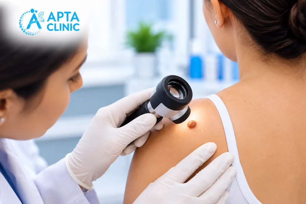
The clinician may assess the mole’s shape, border, colour and surface. A magnifying examination tool may be used when appropriate.
A visual assessment may be sufficient for some typical lesions. If there is uncertainty or concern, the clinician may recommend a biopsy or referral for further evaluation.
Step 3: Discuss the appropriate next step
Depending on the findings, the options may include monitoring, removal or further testing.
If removal is recommended, ask why that method is appropriate, whether the tissue will be examined in a laboratory, what risks are involved and how the wound will be cared for.
Step 4: Confirm the treatment and follow-up plan
Before a procedure, confirm the expected recovery, wound-care instructions, possible scarring, total cost and follow-up arrangements.
If the lesion is being investigated for possible skin cancer, make sure you understand how and when the results will be communicated.
What to Expect During Recovery
Recovery depends on the removal method, the location and size of the wound, and your individual healing.
A small procedure may allow you to resume many normal activities quickly, but the wound still needs appropriate care.
After removal, your clinician may advise you to:
- Keep the wound clean and protected as directed.
- Change dressings according to the instructions provided.
- Avoid picking at scabs or disturbing healing skin.
- Avoid friction, stretching or activities that put pressure on the wound while it heals.
- Use only recommended products on the treated area.
- Attend follow-up appointments and obtain any recommended laboratory results.
Seek medical advice if you notice increasing redness, swelling, pain, pus, persistent bleeding, fever or another symptom that concerns you. These symptoms can indicate a complication and should be assessed.
Follow the specific aftercare instructions provided for your procedure rather than relying on general advice alone.
Will Mole Removal Leave a Scar?
Mole removal can leave a scar, particularly when a surgical procedure creates a wound. The final appearance depends on the removal technique, the mole’s size and location, the way the wound is closed, skin characteristics and individual healing.
Other possible effects include temporary redness, changes in pigmentation, infection or bleeding. Some scars become less noticeable over time, but no procedure can guarantee a scar-free result.
If the mole is being removed for cosmetic reasons, discuss the likely scar before consenting to treatment. The clinician should also explain whether the chosen method could affect the ability to examine the tissue if a diagnosis is needed.
Mole Removal Cost in Mysore
The cost of mole removal in Mysore depends on several factors:
- The size, depth and location of the mole.
- Whether one mole or several moles need treatment.
- The removal technique required.
- Whether local anaesthesia or wound closure is needed.
- Whether a biopsy or laboratory testing is recommended.
- The need for follow-up visits or wound care.
A verified current mole-removal price for APTA Clinic was not provided in the source information for this article. Contact the clinic to ask about the consultation charge, available procedures, laboratory costs where applicable and follow-up fees.
If you have health insurance, check with your insurer about coverage for a medically indicated procedure. Cosmetic removal may be treated differently from removal for diagnostic or medical reasons, depending on the policy.
Choosing a Clinic for Mole Assessment in Mysore
When choosing where to have a mole assessed, consider the clinician’s relevant qualifications, the approach to examining suspicious lesions, access to appropriate diagnostic testing and the clarity of the aftercare and follow-up plan.
Before proceeding, ask:
- Who will assess the mole, and what are their relevant qualifications?
- Does the lesion need further investigation before removal?
- Will the removed tissue be sent for laboratory examination if indicated?
- What are the possible risks and likely scar?
- What will the total cost include?
- How will I receive the results and arrange follow-up?
These questions are particularly important when a mole has changed or appears unusual. A cosmetic procedure should never take the place of appropriate medical assessment.
Mole Assessment at APTA Clinic in Mysore
APTA Clinic is described in the supplied information as a multi-specialty aesthetic clinic in Mysore with a dermatology department. If you are concerned about a mole, contact the clinic to ask whether it can assess your particular concern and whether mole removal is available.
APTA Clinic contact details
- Phone: +91 7411 707 555
- Krishnamurthy Puram: Mysore, Karnataka 570004
- Vijayanagar: Mysore, Karnataka 570030
- Website: https://aptaclinic.com/
Confirm the exact branch address, appointment availability, clinician details and current charges directly with the clinic before visiting.
If a mole is changing, bleeding, persistently itching or otherwise unusual, seek medical assessment promptly rather than treating it as a cosmetic issue.
Frequently Asked Questions (FAQs)
No. Most moles are harmless and do not need removal. A clinician may recommend monitoring, removal or further investigation depending on the mole’s appearance, symptoms and medical history.
Some moles can be removed for cosmetic reasons after appropriate assessment. The clinician should first determine whether the lesion has features that require diagnostic investigation.
The experience depends on the procedure and location. Local anaesthesia is commonly used for surgical removal to reduce pain during the procedure. Some tenderness or discomfort may occur afterward.
A mole can sometimes recur or pigmentation can reappear after certain removal methods. If a growth returns or changes after removal, arrange a medical assessment rather than attempting to remove it again yourself.
Prices vary according to the mole’s size, location, removal method and whether laboratory testing or follow-up is required. Contact APTA Clinic for current charges because a verified price was not available for this article.
A changing mole should be assessed promptly by a healthcare professional. The clinician will decide whether monitoring, biopsy, removal or referral is appropriate. Do not attempt cosmetic removal before the lesion has been assessed.
No. The appropriate method depends on the type of lesion and whether tissue needs to be examined under a microscope. A clinician should determine the safest and most suitable approach.
The supplied information describes APTA Clinic as having a dermatology department, but its dedicated mole-removal service and specific procedures have not been verified. Contact the clinic on +91 7411 707 555 to confirm whether assessment and treatment are available.